Anyone can experience dental tissue damage. If such a problem arises, how a tooth piece has broken off, what to do and who to contact - it is necessary for everyone to know. The loss of a fragment of enamel or dentin, pulp exposure, damage to the mucous membranes of the oral cavity are not all possible consequences, the worst thing is deep infection with the entry of bacterial agents into the systemic circulation and the formation of purulent foci in any organs and tissues.
Content
The reasons
There are many factors that lead to the violation of the integrity of teeth. The most common are:
- Traumatic injuries. Any fall or blow, caused by a heavy object, can lead to chipping.
- Power failure. Frequent consumption of foods high in carbohydrates provokes the appearance of a multitude of microscopic cracks, which later under the influence of too cold or hot food, food with abrasive substances (nuts, caramel, etc.) lead to irreversible loss of tooth tissues.
- High acidity in the mouth. This condition is observed in endocrine and gastrointestinal pathologies (gastritis with increased acidity, peptic ulcer and duodenal ulcer, gastroesoreflux disease), inflammatory lesions of the oral cavity (stomatitis, gingivitis, periodontitis). As a result, the areas of the surface enamel are destroyed by the action of acid and become sensitive to any, even the most insignificant, effects.
- Sharp temperature change. Tooth tissues are sensitive to significant differences and are easily deformed (for example, if you use hot tea after ice).
- Violations of mineral metabolism and lack of vitamins. If the diet is not rich in calcium, phosphorus and other essential salts, then the physiological processes of building and maintaining the integrity of the enamel are disrupted.
- Malocclusion Incorrect tooth position, inadequate installation of fillings and crowns (especially metal ones) create a situation in which certain areas of contact between the upper and lower teeth experience excessive stresses. The result is always the same - erasing tooth enamel.
- Pathology of the teeth. Caries or tartar not only changes the architectonics of an organ, contributes to the formation of potentially dangerous sharp fragments, but also reduces the density of healthy tissues.
- Bad habits. There are many of them: cleaning the seeds with teeth, sucking pens and other hard objects, opening sealed bottles with drinks, etc. Systematic exposure will inevitably lead to multiple destruction of the enamel.
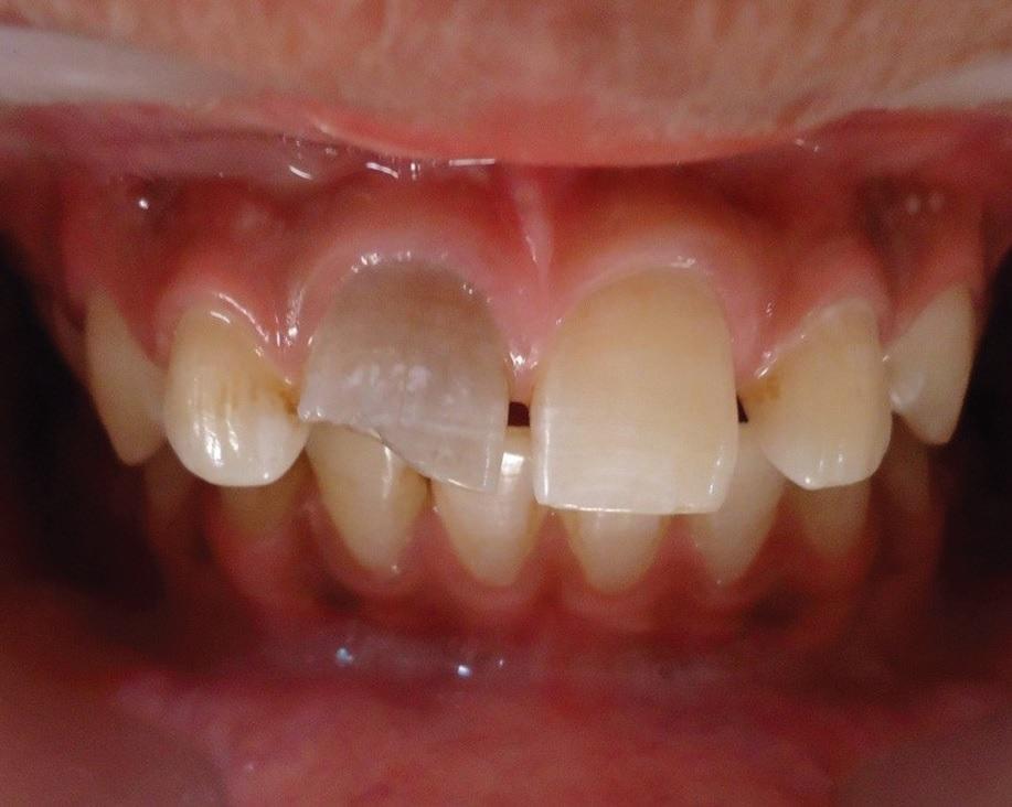
Consequences of accidental hammering
Types of chips
There are several classifications of chipped teeth, but the most common is the gradation of the depth of the lesion. Allocate:
- Incomplete chipping or microcrack. The lesion captures only the surface layers of the tooth; individual pieces or fragments are not separated, but the organ becomes less resistant to future negative influences.
- Chip enamel. Any fragments of tooth enamel are completely lost. Usually, such a condition is noticed by the patient himself at random inspection of the tooth.Given the volume of the surface layer, we can conclude that the front teeth are most susceptible to such troubles (incisors, extremely rarely - fangs).
- Chip dentine - soft cement material. This outburst of the tooth becomes extremely sensitive even to weak loads (consumption of regular food). Fragmentation and crumbling of the dentin can lead to complete loss of a tooth.
- Chip with pulp exposure - loose fibrous connective tissue with many blood and lymphatic vessels, nerve endings. The main danger is the penetration of infection and the possibility of the development of remote purulent complications in the body.
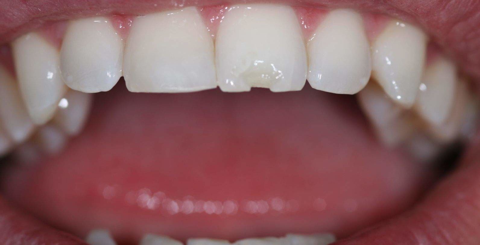
Severe dentin stripping
The first two states are considered non-dangerous and are often not recognized in time, which leads to the further progression of pathology and the development of unpleasant consequences.
Depending on the duration of the impact of the traumatic factor, chipped teeth can be acute or chronic.
The clinical picture of chipped teeth
Manifestations of fractures depend on the specific type. With microcracks clinical symptoms are completely absent, due to the remoteness of the nerve endings.
Chips of tooth enamel are characterized by a slight increase in sensitivity to cold and hot products. Often, the problem is not given due attention, and the defeat is steadily progressing, leading to irreversible consequences from the deeper parts of the masticatory organ.
It is important! Damage to the dentin is more serious. Sensitivity to cold (less than 10–15 degrees) and hot (over 40) products becomes more pronounced. Additionally, there is discomfort and slight soreness towards any sour and sweet substances.
With involvement in the trauma of the pulp, there is a strong pain syndrome. Painful sensations are extremely acute, aggravated by any contact with food or liquid, and increase significantly when exposed to excessively low or high temperatures. The pain is so pronounced that most non-narcotic analgesics do not bring a noticeable improvement in the condition.
Separately, it should be noted features of irradiation - the spread of pain beyond the lesion. The essence of the phenomenon is the innervation of the patient's tooth and other tissues with the same nerve fiber.
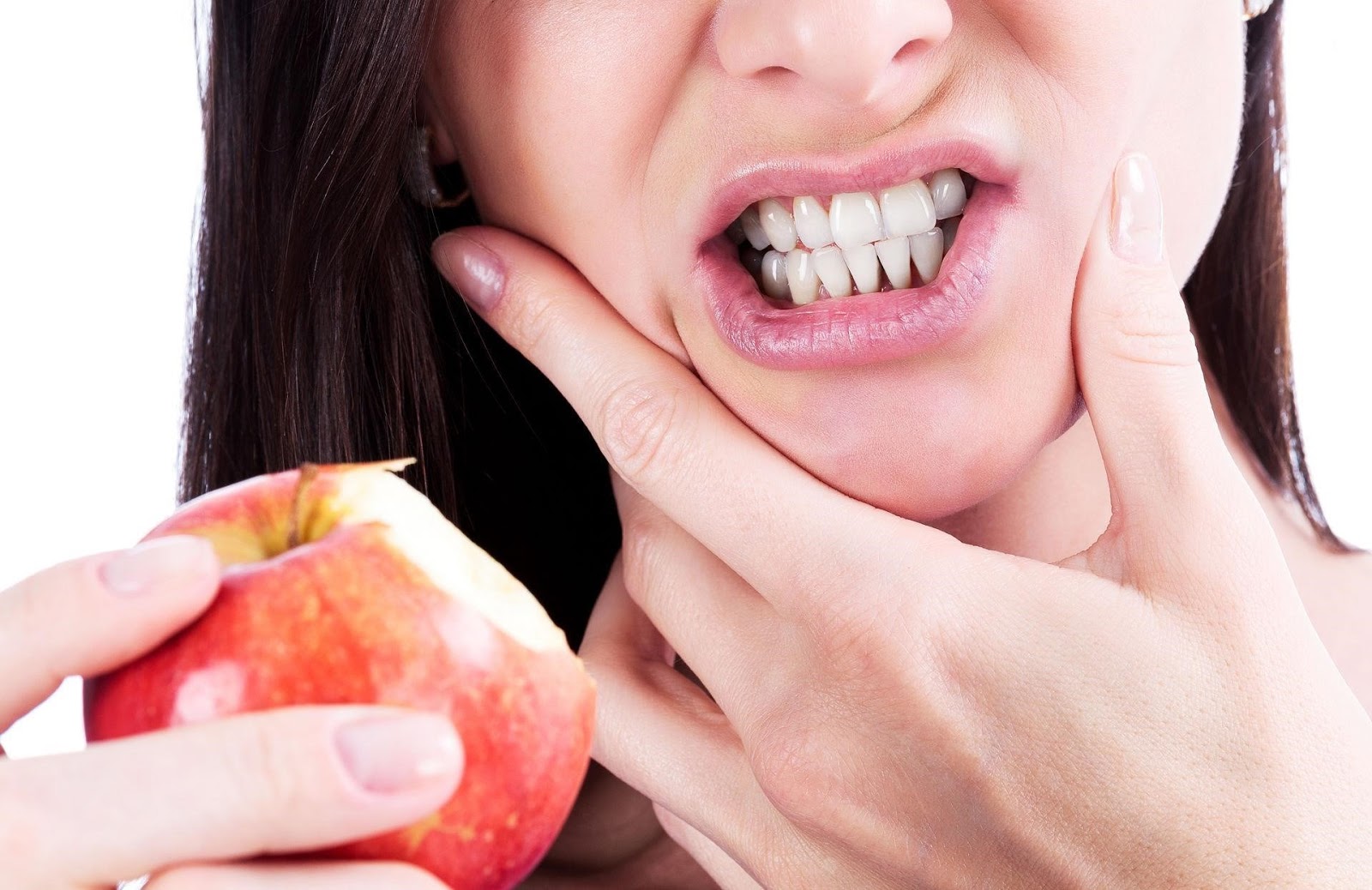
The slightest chipping of the tooth can cause unbearable pain.
The maxillofacial region is controlled by the trigeminal nerve (V pair of cranial nerves), which has three branches - the infraorbital, maxillary and mandibular. Every three pairs are responsible for the innervation of the right and left half of the front of the head, respectively. In the central areas, the nerve trunks form multiple anastomoses (connections), which causes the appearance of cross-pain reactions.
| Localization of the lesion | Radiation Area |
| Upper jaw, incisors and canines. | Brow arc, temporal region. |
| Fangs, premolars, first molars. | Zygomatic arches, mandibular region. |
| The first molar of the upper jaw. | Temple area. |
| 1, 2 and 3 molars of the upper jaw. | Parotid region. |
| The first molar of the lower jaw. | Arch of the mandible, retromandibular region. |
| The third molar of the lower jaw. | Chin, submandibular and parietal region. |
| Cutters, canines. Premolars and first molar of the lower jaw. | Chin area. |
Knowledge of the places of irradiation of pain helps in the differential diagnosis, when identifying the affected area causes significant difficulties.

Areas of irradiation of toothache: 1 - brow arc, 2 - okolonosovaya region, 3 - zygomatic arch, 4 - temporal region, 5 - parotid region, 6 - mandibular joint, mandibular arch, 7 - chin region, 8 - parietal region, 9 - chin.
In case of severe damage to the pulp or gums, bleeding may develop.As a rule, it is insignificant and ends within 3–8 minutes.
First Aid Algorithm
In the case of spalling of a fragment of tooth enamel or deeper tissues, as well as the appearance of cracks:
- To clear an oral cavity from contents (parts of food or liquid). To do this, rinse your mouth with warm water. The ideal temperature is 38–45 degrees.
- Treat the affected area with antiseptics. Hydrogen peroxide 3% or chlorhexidine 1% can be used.
- In case of bleeding, take a cotton swab, moisten it with an antiseptic solution and attach it to the affected area.
- If pulp tissue is exposed, then any movement of air in the mouth (speech or breathing) will cause severe pain. It is necessary to cover the lesion with a dry cotton swab.
- With severe pain, it is allowed to take any non-steroidal anti-inflammatory agent. The strongest is Ketorol (up to 3 tablets per day).
Antiseptic treatment is essential for the prevention of early and delayed infectious complications. Damage to the pulp and mucous membranes of the oral cavity can easily become infected, and active reproduction of the bacterial flora will be observed in the tooth tissues.

After taking painkillers, you should seek help from a specialist.
After first aid it is forbidden to eat any food. Food can not only cause a lot of discomfort (pain), but can aggravate the general condition.
An urgent need to resort to the help of a dentist (preferably immediately). Such treatment is considered in clinics without an appointment.
It is important! Adequate medical care provided in a timely manner will help preserve the integrity of the tooth, prevent the appearance of purulent-septic complications and return to the usual way of life.
What can not be done? In the process of first aid it is strictly prohibited:
- Insert whole or crushed analgesic tablets into the affected tooth and under the gum, as well as dissolve painkillers in the mouth. The drug particles clog up all the affected gaps and only exacerbate the pain syndrome, while the analgesic effect practically does not occur.
- Trying to return the broken-off fragment of the tooth to its former place. In addition to additional injury to hard and soft tissue, there will be no improvement.
- You can not brush your teeth with toothpaste and other personal hygiene products. Such a procedure will be extremely painful, significantly increasing the risk of introducing pathogenic flora into the wound defect.
- Do not delay the visit to the doctor. Even with a slight chipping (small area of enamel), the tooth loses its resistance to the mechanical and chemical factors of aggression that are constantly present in the oral cavity. Chewing movements and speech, as well as food particles and enzymes secreted in the oral cavity, will contribute to the progression of the lesion (deep tissue involvement). The clinical picture, in any case, will become brighter.
Dental restoration
Currently, there are many options to restore the integrity of the tooth. Among them are:
| Method name | Suitable teeth |
| Direct Art Restoration | Cutters, canines. |
| Veneers, Lumineers | Cutters, canines. |
| Tooth Tabs | Molars, premolars. |
| Crowns | Any type of teeth, including dairy |
| Implants | Any teeth (good for replacing the front group) |
Art restoration
The treatment is engaged in a dentist-therapist. The procedure allows to restore the integrity of the tooth enamel, provided that the dentin is not completely damaged, and the pulp is not affected. The method is suitable for restoring the anterior group of teeth (canines and incisors) and is extremely rare for the treatment of others.
At the first visit to the doctor after carrying out infiltrative or conductive anesthesia, nonviable enamel tissue is removed (superficial, within 0.3–1 mm).
Then hardened materials are applied in layers (cements, composites or photopolymers). The thickness of each layer is about 1–2 mm; an artistic correction is immediately carried out (the creation of an anatomical shape and a chewing surface inherent in the tooth prior to the damage, and to adjacent bone organs).
In most cases, the procedure is painless and takes no more than 30 minutes of time.

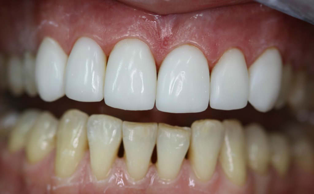
Restoration of chipped tooth by direct restoration
Installation of veneers and lumineers
In addition to composite materials, veneers - ceramic coatings are used to restore the tooth. They are set for visual masking of chips and cracks, as well as color defects (dark spots). The procedure is performed by an orthopedic surgeon, ideal for front teeth.
It is important! Veneers are selected individually depending on the anatomical features of the tooth (according to its cast).
The impressions are made of gypsum, then ceramic plates are stamped in the laboratory according to the models.
Before installation, the tooth is turned (for better fixation). Consequently, the method is extremely traumatic, and after manipulating the veneers you will have to walk all your life, only replacing those that were destroyed or fallen away. When the enamel surface is prepared, veneer is attached to a special glue.
This is how veneers are installed on the front row of teeth.
One of the varieties of veneers are Lumineers - thin (no more than 0.3 mm) ceramic plates that have appeared on the market of medical services in the past few years.
The main feature of lumineers is the absence of teeth turning. Due to the individual selection and high fineness, they are glued to the original tooth without prior preparation.
Tooth Tabs
Dental tabs are a large filling that is not made in the patient’s mouth, but in the dental laboratory after taking individual impressions. A distinctive feature is the perfect match to the anatomical surfaces of the tooth, providing a tight joint.
The tab can restore only the crown of the tooth, provided that it is destroyed by no more than 1/3.
It is important! The front rows of teeth cannot be restored by this method, only molars and premolars.
Tabs are of two main types:
- Composite. Such materials are significantly inferior in strength metal, but never subside. They do not break the bite and do not deform the act of chewing.
- Metallic. Currently, the method is obsolete in most clinics, since these tabs have several disadvantages: poor adhesion to the tooth tissues, the possibility of galvanic reactions due to the effects of currents (increased salivation, burning sensation, etc.).

Tooth metal tab mounted on the tooth layout
In most cases, dentures are placed on the teeth with a live neurovascular bundle. The procedure takes place in several stages:
- Dissection A tooth is ground to remove damaged tissue.
- Plaster casting.
- Making tabs (usually up to 7 days).
- Fixation. The dental surface is given a rough look (with the help of sandblasting), then composite glue is applied to it and the filling material is fixed.
Dental crowns
Crowns are shown with significant damage to the tooth (more than 2/3). Metallic and plastic materials are used. Plastic crowns are much cheaper and inferior in strength. Metal cast on individual casts, then covered with a thin layer of porcelain in order to impart an aesthetic component.
Installation can be made on all teeth (incisors, canines, molars, premolars).
The procedure can be represented as follows:
- Treatment of a destroyed tooth. Removed non-viable tissue, pulp, dental nerve. After you install a seal or stump pin tab.
- Plaster casting.
- Making a crown on the plaster pad.
- Fixing the crown with cement.

Crown installation process
There are certain types of crowns for children, which are installed on baby teeth and standing molars. As a rule, they are not made on individual casts. Thanks to the thinnest walls, crowns are easily fitted to the dental surfaces. Fastening occurs with the use of composite glue.
Implantation
This method is ideal for small lesions (1–2 teeth) of the front teeth (incisors, canines).
After complete removal of the tooth (2–3 weeks) at the stage of preparation for treatment, the dentist studies the condition of the jaw bone tissue, determines the viability of the adjacent teeth areas (using panoramic X-ray or computed tomography).
If the bone skeleton is intact, then you can proceed to the procedure, if the bone volume is insufficient, the procedure for its extension is carried out (implantation of artificial material or bone block), after which a long recovery period is required - from 120 to 180 days.
It is important! Implant placement is a complicated dental operation. It is performed under conduction anesthesia.
The course of the operation can be represented as follows:
- Dissection of the gums with a scalpel or laser to create the necessary surgical access.
- Drilling holes in the bone tissue and removing unnecessary fragments.
- Installation in the created hole of the dental implant. Then it becomes covered with bone chips and a cap.
- If necessary, the artificial formation of the gums (allows you to create a smooth edge, increase the volume of the mucous membrane for a firm fixation and prevention of the ingress of food and fluid particles in the future).
- Closure of the gums.
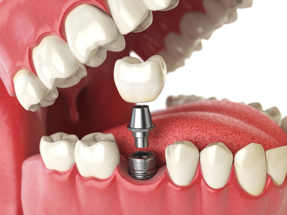
Installation scheme of an implant with an abutment and a crown
Upon completion of the manipulations, a period of engraftment is required - from 3 to 5 months. At this time, it is necessary to visit the doctor every 2–4 weeks to monitor the condition of the new tooth. To preserve the aesthetic appearance of the teeth, a butterfly cap is installed, which clings to the adjacent teeth without injuring them.
After 90–150 days from the moment of implantation, the cap is removed, instead of it an abutment is attached - a cone-tip, on which the prosthesis will be fixed. There are 2 options for mounting the prosthesis:
- fixed (installed on a specialized glue);
- removable (fastening is carried out on a screw basis).
In the rehabilitation period (3-4 days after each intervention) can be observed:
- edema;
- slight or severe pain;
- bleeding.
Video: what to do if a tooth broke
Cost of tooth restoration
The price for the provision of services for the restoration of teeth varies significantly depending on the type of dental clinic (public or private), region and materials used in the course of manipulation. The minimum cost is as follows:
| Recovery Method Name | Price |
| Art restoration (direct) | About 2000 rubles |
| Veneers | From 8,000 rubles |
| Lumineers | From 30,000 rubles |
| Tooth Tabs | From 6000 rubles |
| Installation of crowns | From 10,000 rubles |
| Dental implantation | From 20,000 rubles |
Complications
In addition to many unpleasant subjective sensations and violations of the quality of life, can develop:
- Infection of the pulp. Bacterial and viral agents penetrate deep into the internal tissues, leading to the development of an inflammatory process. As a result, the tooth is rapidly destroyed in all directions from the center, there may be distant foci - abscesses and cellulitis of the gums, jaw tissues, less often - septic foci in any organ.
- Formation of cysts and granulomas. Infection of tissues, failure of local factors of immunity, violation of the anatomy of the tooth provokes these pathologies.
- The change in the angle of inclination of the tooth with a violation of bite and traumatic damage to other masticatory elements. In some cases, possible fragmentation of the jaw bone.
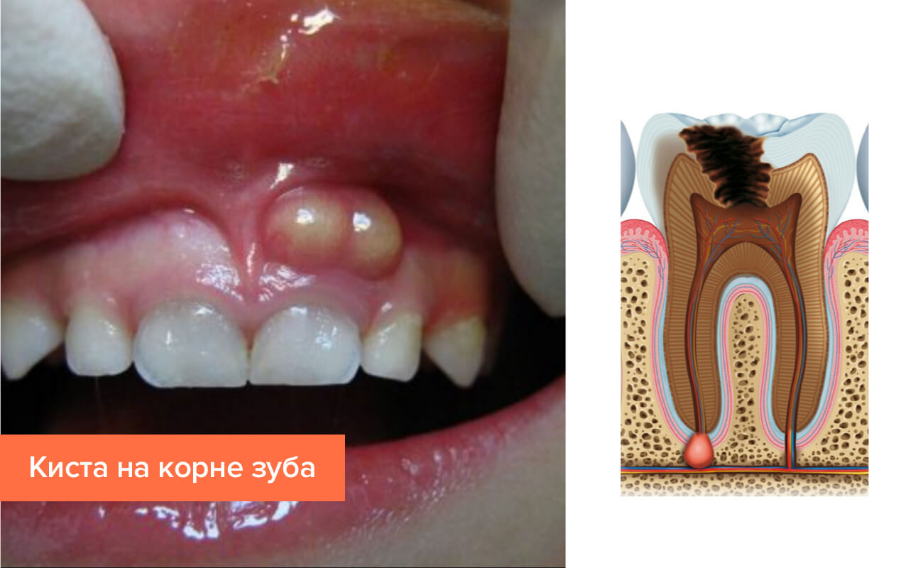
Formation of a cyst is one of the possible complications when a tooth is chipped
Thus, such a problem as chipping a part of a tooth is a serious one.The patient experiences many unpleasant feelings (pain, hypersensitivity to cold or hot, sour or sweet foods), and also runs the risk of serious surgical pathology - abscesses, cellulitis, etc. since there is a great opportunity for the development of complications, which significantly increase the duration of disability and the cost of treatment.


